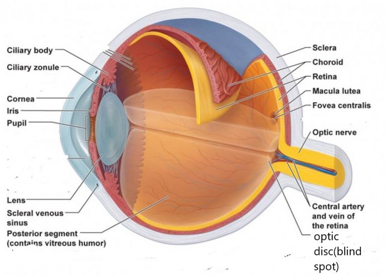
An intact epithelium is necessary for maintenance of corneal transparency. The regenerative capability of injured corneal epithelium is pronounced mitotic divisions, along with cell movements, ensure a rapid return of injured epithelium to normal structure.
VASCULAR TUNIC FREE
Numerous free nerve endings are present among the epithelial cells.

The numerous microvilli on the surface of the superficial cells function to retain the tear film on the corneal surface. The epithelial cells are tightly packed, interdigitate profusely, and adhere through numerous desmosomes. Large animals have more layers of polyhedral and squamous cells. In the dog, cat, and bird, the corneal epithelium consists of a single layer of basal cells, two to three layers of polyhedral (wing) cells, and two to three layers of nonkeratinized squamous cells. The anterior (corneal) epithelium is nonkeratinized stratified squamous, between 4 and 12 layers in thickness ( Fig. The cornea is composed of five layers: (a) anterior epithelium, (b) subepithelial basement membrane, (c) substantia propria, or stroma, (d) posterior limiting lamina (Descemet’s membrane), and (e) posterior epithelium (corneal endothelium). The horizontal corneal diameter is greater than the vertical corneal diameter, resulting in a mildly elliptical shape. Because the cornea also has a radius of curvature smaller than that of the sclera, it is more curved than the sclera. The transparent cornea is a convex–concave lens, slightly thicker at the periphery than at the center, and with a smaller radius of curvature centrally than peripherally. The optic nerve leaves the eye through numerous perforations in a sievelike area of the sclera referred to as the area cribrosa. A firm attachment to the sclera is provided for the tendons of the extraocular muscles through the interweaving of tendon and scleral fibers. The orbital fasciae, located outside the sclera, consist of the periorbita (connected to the external or dural sheath of the optic nerve), the bulbar sheath (Tenon’s capsule), and the fascial sheaths of the extraocular muscles. In the layer of the sclera adjacent to the choroid, elastic fibers predominate, and fibroblasts and melanocytes are more numerous this layer is referred to as the lamina fusca sclerae. These fibers are arranged meridionally, obliquely, and radially in an irregular pattern.

The sclera contains primarily collagen fibers but elastic fibers, fibrocytes, and melanocytes (anteriorly) are interlaced among the collagen fibers. The sclera is thickest at the limbus (junction of the cornea and sclera) in dogs and cats and in the region of the optic nerve in ungulates. The sclera is thinnest at the equator of the globe. Thickness of the sclera varies in different parts of the eye and among species.

Some ungulates, such as the horse and large ruminant, have globes that are slightly compressed in the anteroposterior axis. While the size of the globe can be quite variable in different species, the shape is essentially spherical. The sclera is a layer of dense irregular connective tissue that protects the eye and maintains its form (shape).


 0 kommentar(er)
